Unlock High-Throughput Cellular Insights with Advanced High-Content Screening Systems
Scalable, Automated Solutions for Precise Cellular Phenotyping and Mechanism-of-Action Studies.
Uncover Complex Cellular Phenotypes with High-Content Screening Platforms
High-Content Screening (HCS) combines automated microscopy with powerful image analysis software to extract detailed quantitative data from cellular assays. By enabling multiplexed measurements of morphology, fluorescence intensity, and subcellular localization, HCS systems are essential for phenotypic screening, mechanism-of-action studies, and compound profiling.
At Cellular Tissue Analysis, headquartered in Indianapolis, IN, our HCS solutions are engineered for scalability, reproducibility, and integration into high-throughput environments. Whether in pharma R&D or academic research, our systems help users gain deep insights from high-volume cellular datasets.
Core System Components for Scalable Phenotypic Analysis
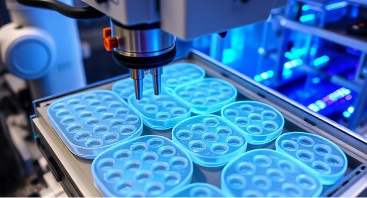
Imaging & Detection Hardware
- Automated fluorescence microscopy with multi-channel imaging:
Optical & Imaging Sensors– enable multiplexed fluorescence capture for high-content cellular screening and phenotyping. - High-resolution CMOS or sCMOS cameras:
Biometric and Health Sensors – deliver sensitive, low-noise image detection for precise quantification in live-cell imaging. - Environmental control for live-cell imaging (CO₂, temp, humidity):
Environmental & Agriculture Sensors – maintain stable conditions to support cell viability during long-term time-lapse assays.
Software & Image Analytics Tools
- Machine learning–based image segmentation
- Feature extraction for cell count, shape, intensity, localization
- Multiparametric assay scoring
- Batch processing and statistical clustering
- Customizable analysis pipelines with AI-assisted annotation
Cloud & Informatics Integration
- Data upload to secure cloud repositories
- API integrations with LIMS, ELN, and compound libraries
- Remote review, QC, and result sharing
- Collaborative dashboards and searchable image archives
- Scalable storage for large image datasets
Notable Features
Screens hundreds of wells per hour
Multi-parametric output from single experiments
Supports 2D, 3D, and organoid assays
Compatible with wide range of fluorescent dyes and biosensors
Automated plate handling for HTS environments
Integration Capabilities
- Full integration with robotic screening workflows
- Synchronization with liquid handling and compound libraries
- Compatible with AI/ML-based hit identification platforms
- Data transfer to cheminformatics and omics pipelines
- Remote monitoring and lab automation support
Applications Across Life Science Disciplines
- Drug target validation and lead optimization
- Cytotoxicity and genotoxicity profiling
- Cell cycle and apoptosis analysis
- Stem cell differentiation and morphology assays
- Host-pathogen interaction studies
Why Choose Our HCS Systems
- Combines speed and image depth for large-scale analysis
- Reduces false positives via multiparametric validation
- Enables unbiased phenotypic discovery
- Facilitates mechanism-of-action studies
- Scalable from 96- to 1536-well formats
Standards & Compliance
- GxP-compliant platforms with audit trails
- 21 CFR Part 11–ready for regulated workflows
- Integration with ISO and OECD test guidelines
- CE-marked systems for international compliance
- Automated calibration and performance validation tools
Industries & User Groups
Pharmaceutical R&D and Screening Labs
Biotechnology and Biologics Development
Academic Translational Research Centers
Contract Research Organizations (CROs)
Toxicology and Preclinical Testing Facilities
Case Studies
Digitally transform your cell screening capabilities with advanced HCS technologies.
Contact Cellular Tissue Analysis for personalized solutions or a system demonstration.