Unlock Gene Expression Insights with Spatially
Resolved Transcriptomics
Reveal Spatial Gene Expression with Precision—Integrate Tissue Morphology and Transcriptomic Insights

Uncover Molecular Landscapes with Spatial Transcriptomics
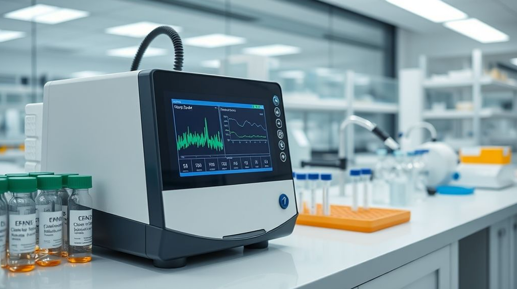
Spatial Transcriptomics Platforms enable the precise mapping of gene expression across tissue sections, preserving spatial context while capturing transcriptomic data. These technologies revolutionize how researchers understand tissue heterogeneity, cellular interactions, and disease microenvironments.
At Cellular Tissue Analysis, based in Indianapolis, IN, our platforms combine high-throughput sequencing, imaging, and advanced informatics—delivering an integrated workflow for spatially resolved transcriptomic profiling in both research and clinical labs.
Core Components of Spatial Transcriptomics Systems
Hardware
- Spatially barcoded glass slides or arrays:
Optical & Surface Sensors – detect spatial positions and barcodes on the array to preserve tissue context during imaging and sequencing. - Capture spots with oligo-dT or targeted probes:
Hybridization Detection Sensors – confirm probe-target binding through fluorescence or chemiluminescence-based detection modules. - Compatibility with FFPE and fresh frozen samples:
Temperature & Humidity Sensors – ensure preservation conditions for sample integrity across processing types. - Brightfield and fluorescence microscopy integration:
Imaging Sensors (CCD/sCMOS) – support multi-modal data capture for morphology and molecular signal overlay. - Hematoxylin & Eosin (H&E) or IHC co-staining capability:
Spectral Discrimination Sensors – distinguish staining components via multispectral or colorimetric detection.
Spatial Informatics Software
- Gene expression heatmap visualization
- Tissue segmentation and cell-type annotation tools
- Multi-omic overlay (e.g., transcriptomics + proteomics)
Molecular Processing & Sequencing Prep
- On-slide reverse transcription and cDNA generation
- Indexed library preparation workflows
- Compatibility with NGS platforms (e.g., Illumina, Oxford Nanopore)
Key Features
Subcellular to multi-cell resolution options
High transcript capture sensitivity
Full-tissue section coverage or ROI-based mapping
Compatibility with clinical pathology workflows
Open-source and proprietary software options for analysis
Why Choose Our IHC Stainers
- Accelerated turnaround for diagnostic results
- High reproducibility across large sample volumes
- Reduces technician workload and hands-on time
- Built-in waste tracking and reagent economy modes
- Supports a broad range of biomarkers and chromogens
Application Areas
- Cancer biomarker detection (e.g., HER2, Ki-67, PD-L1)
- Neurodegenerative and infectious disease pathology
- Tissue-based drug discovery and target validation
- Transplant histocompatibility assessments
- Immunophenotyping in veterinary pathology
Integration Capabilities
- Full integration with pathology LIS systems
- Compatibility with digital slide scanners for post-stain imaging
- Remote access and protocol editing via secure networks
- Workflow optimization through AI-based stain intensity analysis.
Regulatory & Compliance Standards
- CE-IVD and FDA-cleared (where applicable)
- ISO 13485 certified for medical device quality
- Compatible with CAP and CLIA-accredited workflows
- 21 CFR Part 11–ready for digital recordkeeping
- Validated protocols for diagnostic-grade biomarker testing
Industries We Serve
Cancer Research and Precision Oncology Labs
Academic Genomics and Neuroscience Institutes
Pharma R&D for Biomarker and Drug Target Discovery
Pathology Departments Exploring Molecular Diagnostics
Multi-Omics Core Facilities
North American Case Studies
Harness the power of location-specific gene expression.
Connect with Cellular Tissue Analysis for customized platform demonstrations, pricing, and implementation support.
