Isolate Cells with Surgical Precision
Non-contact systems designed for reliable, high-fidelity molecular analysis.
Precision Cell Isolation for Molecular Clarity
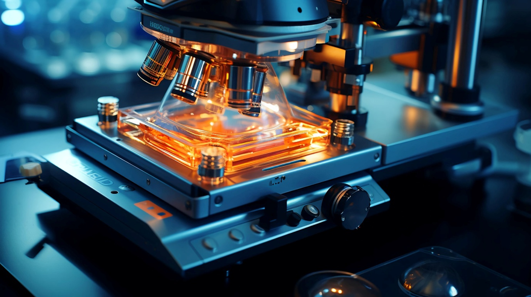
Laser Capture Microdissection (LCM) Systems empower researchers to isolate specific cells or microscopic regions from tissue sections under direct visualization. This enables high-fidelity molecular analysis—by eliminating cellular noise and ensuring sample purity.
At Cellular Tissue Analysis, headquartered in Indianapolis, IN, our LCM platforms combine high-resolution optics, targeted laser control, and contamination-free collection systems, enabling researchers to retrieve intact, analyzable biomolecules from defined tissue microenvironments.
Core Components of LCM Systems
Hardware
- Spatially barcoded glass slides or arrays:
Optical & Surface Sensors – detect spatial positions and barcodes on the array to preserve tissue context during imaging and sequencing. - Capture spots with oligo-dT or targeted probes:
Hybridization Detection Sensors – confirm probe-target binding through fluorescence or chemiluminescence-based detection modules. - Compatibility with FFPE and fresh frozen samples:
Temperature & Humidity Sensors – ensure preservation conditions for sample integrity across processing types. - Brightfield and fluorescence microscopy integration:
Imaging Sensors (CCD/sCMOS) – support multi-modal data capture for morphology and molecular signal overlay. - Hematoxylin & Eosin (H&E) or IHC co-staining capability:
Spectral Discrimination Sensors – distinguish staining components via multispectral or colorimetric detection.
Molecular Processing & Sequencing Prep
- On-slide reverse transcription and cDNA generation
- Indexed library preparation workflows
- Compatibility with NGS platforms (e.g., Illumina, Oxford Nanopore)
Spatial Informatics Software
- Gene expression heatmap visualization
- Tissue segmentation and cell-type annotation tools
- Multi-omic overlay (e.g., transcriptomics + proteomics)
Key Features
Contact-free isolation—no contamination risk
Capture of single cells or multi-cell regions
Compatible with frozen and FFPE tissue typesing
Maintains integrity of RNA, DNA, and proteins
Compatible with downstream NGS, RT-PCR, or proteomics
Integration Capabilities
- Seamless data transfer to NGS pipelines
- Image overlay tools with digital pathology viewers
- API support for bioinformatics platforms (R, Python, Seurat, etc.)
- Integration with spatial proteomics and single-cell datasets
Applications
- Oncology: Tumor microenvironment and immune cell profiling
- Neuroscience: Spatial mapping of brain region transcriptomes
- Developmental Biology: Gene expression in embryonic tissues
- Infectious Disease: Pathogen–host interaction zones
- Regenerative Medicine: Tissue remodeling and cell fate mapping
Benefits of Our Spatial Transcriptomics Platforms
- Reveal tissue architecture alongside gene activity
- Identify spatial biomarkers and disease niches
- Support spatially targeted drug discovery
- Improve understanding of tumor heterogeneity
- Enable multi-modal tissue analysis with histology alignment
Standards & Regulatory Considerations
- Compatible with CLIA/CAP-accredited sample workflows
- Validated protocols for FFPE and fresh-frozen tissues
- Data traceability for 21 CFR Part 11 compliance
- Alignment with MIAME and spatial transcriptomics reporting standards
- Data export options for repositories (e.g., GEO, ArrayExpress)
Industries We Serve
Molecular Pathology & Histology Labs
Academic Medical Research Centers
Cancer Genomics & Immuno-oncology Research
Pharma/biotech R&D for biomarker validation
Clinical Translational Research Programs
North American Case Studies
Get Started
Experience the power of precision isolation. Contact Cellular Tissue Analysis to schedule a demo or receive tailored quotes and workflow recommendations for your LCM needs.