Imaging Flow Cytometers for Quantitative Cell Imaging in Flow
Unite the speed of flow cytometry with the detail of microscopy.
Visualize and Quantify Cells Simultaneously with Imaging Flow Cytometers
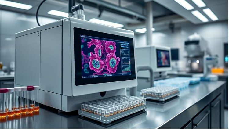
Imaging flow cytometers revolutionize single-cell analysis by merging the high-throughput capabilities of traditional flow cytometry with high-resolution image capture. These instruments enable simultaneous measurement of fluorescence intensity and cellular morphology, empowering researchers to visualize rare events, study subcellular localization, and verify complex gating strategies in real time.
At Cellular Tissue Analysis, based in Indianapolis, IN, we offer next-gen imaging cytometers that support a broad range of cell-based applications—from immune synapse analysis to nuclear translocation and cell cycle profiling.
Core Components: Optical Imaging Meets Cytometric Precision
In addition to our proprietary systems, we support live-cell platforms from global leaders like GAO Tek Inc. and GAO RFID Inc.—combining precision optics with automation-ready configurations.
Hardware Systems
- High numerical aperture objectives for crisp subcellular resolution:
Optical & Imaging Sensors – offer enhanced resolution and light-gathering efficiency to resolve fine structural details in live or fixed cell imaging applications. - Precision fluidics with hydrodynamic focusing:
Motion & Position Sensors – enable consistent cell alignment for accurate interrogation and sorting by tightly controlling sample and sheath fluid dynamics. - Automated sample handling for 96- and 384-well formats:
Motion & Position Sensors – streamline high-throughput analysis with robotic plate loading and precise positioning, ideal for drug screening and multi-well assay workflows.
Software & Analysis Tools
- Morphological feature extraction (size, shape, texture)
- Co-localization and translocation analysis
- Customizable classifiers and machine learning models
- Real-time image capture and gating overlays
- Batch processing and high-content data output.
Cloud/Data Capabilities
- Export to FCS + image files (TIFF, JPEG, etc.)
- Remote collaboration with secure image repositories
- Integration with AI/ML pipelines for phenotype prediction
- Seamless compatibility with image analysis software (IDEAS, CellProfiler)
- Live dashboards for multi-instrument coordination
Standout Features
Capture 10,000+ images per second with real-time classification
Multiparameter imaging across multiple fluorescence channels
Simultaneous brightfield and fluorescence visualization
Robust tools for nuclear/cytoplasmic segmentation and tracking
Exportable annotated image datasets for training AI models
Seamless Integration & Automation-Ready Design
- Compatible with cell culture prep and automated staining systems
- Connectivity with genomic/proteomic profiling pipelines
- Integration with digital pathology platforms
- Automated QC and maintenance alerts via cloud-based dashboards
- API support for custom informatics workflows
Key Advantages
- Deep phenotyping and spatial localization at single-cell resolution
- Visual confirmation of gating and marker expression
- Ideal for rare cell detection and intracellular signaling studies
- Reduces false positives from traditional flow cytometry
- Validated for fixed, live, and apoptotic cell analysis
Research & Clinical Applications
- DNA damage and repair monitoring (γ-H2AX, comet assays)
- Immune cell activation and functional assays
- Nuclear translocation (e.g., NF-κB, STAT) tracking
- Cancer cell identification and drug response visualization
- Apoptosis, necrosis, and autophagy analysis.
Certifications & Standards
- 21 CFR Part 11–compliant for clinical image documentation
- ISO 13485–ready for regulated medical device workflows
- CE-marked instruments for European diagnostic use
- Validated protocols for GLP/GMP environments
- Health Canada–approved systems for research use
Industries & User Segments
Translational Research Institutes
Immuno-oncology and Stem Cell Labs
Biotech and Pharmaceutical R&D Divisions
Biotech and Pharmaceutical R&D Divisions
Academic Cell Biology Departments
Case Studies
Bring visual power to your cytometric workflows with our imaging-enabled platforms.
Contact our experts to schedule a personalized system demo or consultation.